Femoroacetabular impingement (FAI) and hip dysplasia are the two primary causes of hip degeneration, leading to total hip arthroplasty (THA) as the final solution. Hip-preserving surgeries yield worse outcomes when the condition is detected late or significant degeneration has occurred. Additionally, the number of THAs performed in OECD countries is projected to increase substantially by 2050. Reading the large amount of radiographs associated with this poses several problems to orthopedists and radiologists:
HIPPO™ presents a solution that enhances efficiency when reading pelvis radiographs. It significantly reduces daily workload for radiologists reading hip and pelvis x-rays, ensuring consistent and precise reporting. This helps eliminate subjectivity, especially for radiologists who read a high volume of x-rays daily. Furthermore, HIPPO's structured output streamlines the process and aids in identifying changes over time with a standardized methodology.
Total Hip Arthroplasties by 2050 [3]
HIPPO's™ accuracy in measuring angles is within 1 degree
reduction of reading and reporting time when using HIPPO™ [2]
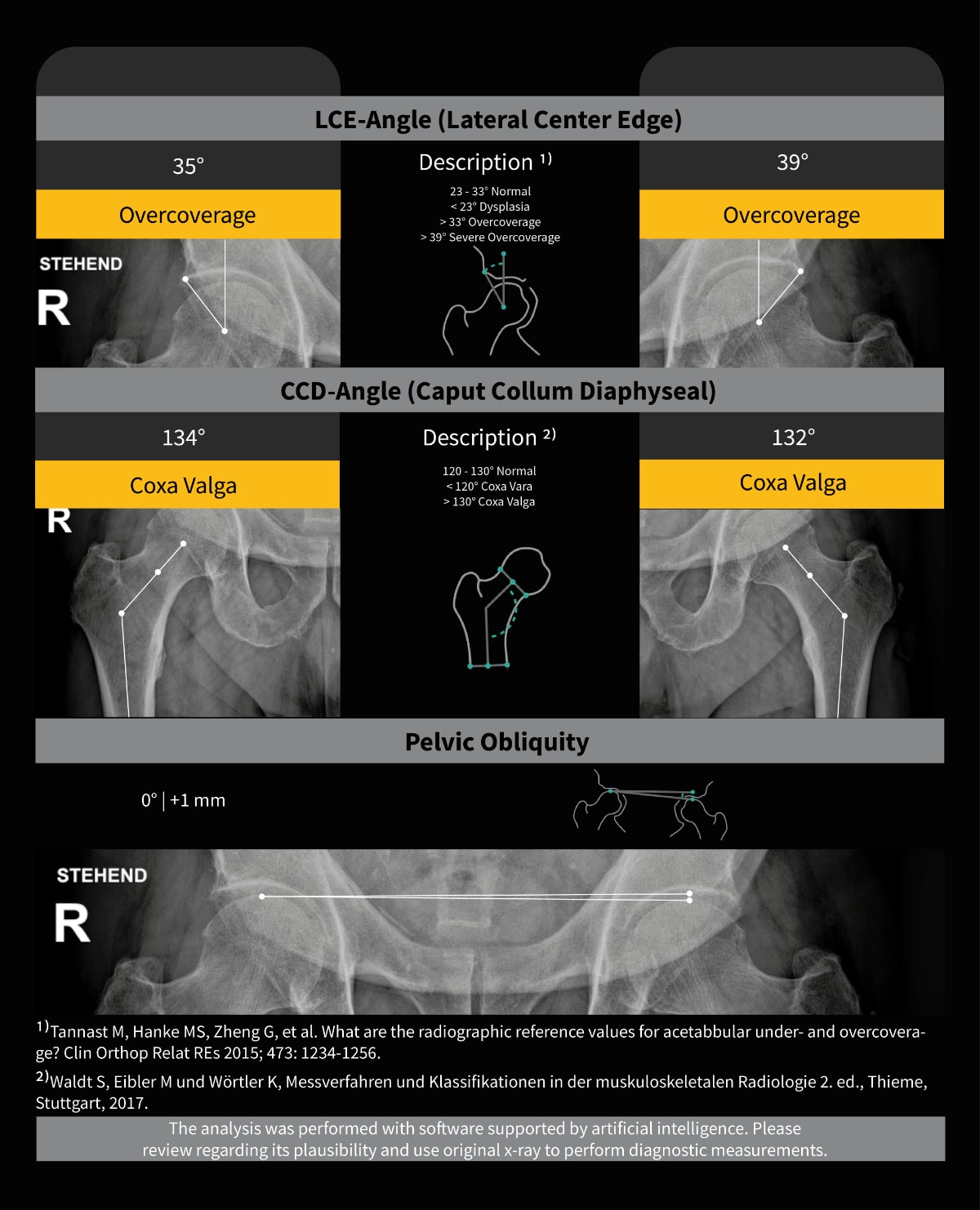
HIPPO™ supports the objective and standardized measurement of the most important hip angles based on digital x-rays. These include, for example, the CCD and LCE angles as well as numerous other relevant angles.
IB Lab HIPPO™ is a radiological fully-automated image processing software device of either computed (CR) or directly digital (DX) images of the pelvis or hip. On either standing or supine, unilateral or bilateral AP radiographs, IB Lab HIPPO is intended to aid medical professionals in the measurement of the Caput-Collum Diaphysis (CCD) angle (also known as Neck-Shaft angle), and the Lateral Center-Edge Angle (LCE), Acetabular Index (aka Acetabular Inclination, Tönnis Angle), Acetabular Angle (aka Sharp’s Angle) and the Femoral Head Coverage/Extrusion Index. On bilateral standing AP hip radiographs, IB Lab HIPPO aids in the detection of the presence or absence of pelvic obliquity.
It should not be used in-lieu of full patient evaluation or solely relied upon to make or confirm a diagnosis.
The system is to be used by trained professionals including, but not limited to, radiologists and orthopedics.
The HIPPO™ Module is intended to be used on adult humans between 18-95 years presenting with hip pain, suspected congenital diseases, femoral-acetabular impingement, spinal malformation or osteoarthritis of the hip.


ImageBiopsy AI software is highly accurate and efficient within our PACS system, which provides valuable information on the status of the knee along the continuum of chondrosis to arthrosis.


The integration of the AI solutions by ImageBiopsy Lab into our RIS and PACS is easy and well done. It is fun to work with and the clarity of the visualized report is an ideal support for our patient consultation.

AI-based solutions reduce the amount of work and the findings become more accurate. An objective value is given which can be used both for monitoring and forecasting the progress. We offer something that others don’t have.

Exact diagnosis and reproducible follow-up exams are indispensable for a successful osteoarthritis therapy. Software-based methods can assist the physician in the therapy management and adjustment process.


